
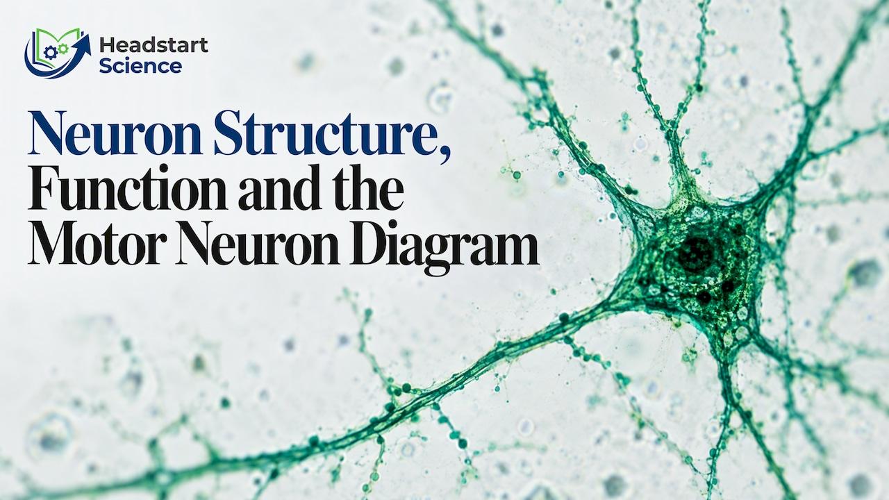
A neuron, or nerve cell, is a cell specialised to carry information as electrical signals around the body. It has a cell body, branching dendrites that receive signals and a long axon that sends them on. Neurons link receptors, the brain and spinal cord, and muscles or glands, so you can sense something and respond in a fraction of a second. This guide covers the parts of a neuron, the three types you meet at GCSE, how to draw and label a motor neuron, and how a signal crosses the gap to the next cell.
TL;DR
- A neuron is a nerve cell that carries electrical impulses. Its three key parts are the dendrites (receive), the cell body (controls the cell) and the axon (transmits).
- GCSE Biology names three types: sensory neurones (from receptors), relay neurones (inside the central nervous system) and motor neurones (to muscles or glands).
- Neurons do not touch. A chemical messenger, a neurotransmitter, carries the signal across each synapse.
- Neurons send and receive signals. Glial cells support them, so a neuron is not the same thing as the nervous system.
- For a motor neuron diagram, draw the pathway in order: receptor, sensory neurone, relay neurone, motor neurone, effector.
Table of Contents
- What is a neuron and what do nerve cells do?
- The parts of a neuron and what each one does
- The three types of neurone at GCSE
- How to read and draw a motor neuron diagram
- How a nerve impulse travels
- The synapse: how the message crosses the gap
- Neuron, glial cell, nervous system: clearing up the confusion
- Exam technique for neuron questions
- Where students usually lose marks
- How we teach the nervous system
- Get live help with GCSE Biology
- FAQ
- Sources
What is a neuron and what do nerve cells do?
Neurons are the cells that do the communicating in your nervous system. The US National Institute of Neurological Disorders and Stroke describes three broad jobs: sensory neurons carry information from the sense organs, motor neurons carry messages to the muscles, and interneurons connect the sensory and motor neurons.

In the UK exam courses the word is usually spelled "neurone", but it means the same thing. Both spellings turn up in textbooks, so recognise each of them.
The reason neurons matter for GCSE is that they explain how an organism reacts to its surroundings. Light, sound, odour, taste, pressure and heat are all stimuli, and sensory neurons detect them. Motor neurons then carry the instruction from the brain to the muscles. Everything from pulling your hand off a hot pan to kicking a ball is a chain of these cells passing a signal along.
The topic sits in the coordination and control part of the course. The OCR GCSE Biology delivery guide lists the central nervous system, sensory and motor neurones, sensory receptors and the structure and function of the reflex arc as content to be studied. Other boards phrase it differently but cover the same ground.
The parts of a neuron and what each one does
A typical neuron has three parts you must be able to label and explain. The National Institute of General Medical Sciences gives a clear summary: dendrites receive signals, the cell body contains the nucleus and controls the cell's actions, and the axon transmits messages. The Queensland Brain Institute agrees, describing dendrites as the receiving part, the axon as the transmitting structure and the soma, or cell body, as the part containing the nucleus.
| Part | What it does | Easy way to remember it |
|---|---|---|
| Dendrites | Receive incoming signals | Dendrites are the "doorway" in |
| Cell body (soma) | Holds the nucleus and controls the cell's activities | The control centre |
| Axon | Carries the impulse away from the cell body | The long "wire" out |
| Axon terminals | Release neurotransmitter at the synapse | The "sending end" |
| Myelin sheath | Fatty insulation that speeds up transmission | The coating on the wire |
On the myelin sheath: the Caltech Science Exchange notes that myelin accelerates transmission along the axon. That is why it often appears in exam answers about why some impulses travel faster than others.
Why neurons are shaped the way they are
Exam questions often ask you to link structure to function. The same logic works for almost every part:
- Long axon: it lets a single cell carry a signal over a long distance, for example from the spinal cord towards a muscle.
- Branched dendrites: they give a large surface area, so the cell can connect with many other cells.
- Myelin insulation: it makes transmission faster.
- Nucleus in the cell body: like other cells, the neuron needs genetic instructions to build proteins and stay alive.
When a mark scheme asks you to "explain how the structure is adapted", write the feature, then the effect, then the benefit to the organism. "The axon is long, so one neurone can carry the impulse a long way, so the response is fast." That three-step pattern earns the marks.
The three types of neurone at GCSE
You need to tell sensory, relay and motor neurones apart and place each one in a pathway.
| Type | Carries impulses | Where you find it |
|---|---|---|
| Sensory neurone | From a receptor towards the central nervous system | Between sense organs (such as skin or eye) and the spinal cord or brain |
| Relay neurone | Between sensory and motor neurones | Inside the central nervous system (brain and spinal cord) |
| Motor neurone | Away from the central nervous system to an effector | Between the spinal cord or brain and a muscle or gland |
The NINDS wording for relay neurones is "interneurons", which are the cells that connect sensory and motor neurons. If a question uses the word interneuron, it means the same cell as a relay neurone.
An effector is whatever carries out the response. Electrical impulses travelling along a motor neurone tell a muscle to contract or a gland to release a hormone. A receptor is the sensing end of the pathway, such as a pain receptor in the skin.
How to read and draw a motor neuron diagram
A motor neuron diagram usually shows one of two things: the structure of a single motor neurone, or the whole pathway from stimulus to response. Examiners may ask for either, so practise both.
Drawing a single motor neurone
A motor neurone has its cell body at one end, with short dendrites around it and a very long axon stretching away towards the effector. Follow this order:
- Draw a rounded cell body and put a small circle inside it for the nucleus.
- Add several short, branching dendrites coming off the cell body.
- Draw one long axon leaving the cell body, as two parallel lines.
- Mark the myelin sheath as segments wrapped around the axon.
- Finish with the axon terminals at the far end, next to a muscle fibre or gland.
- Add an arrow showing the direction of the impulse: cell body to axon terminals, towards the effector.
Label each part with a ruled line that touches the structure. Do not let label lines cross each other, because a muddled diagram can lose marks even when you know the content.
Reading the full pathway
The pathway diagram shows the signal moving in a fixed sequence:
Stimulus → receptor → sensory neurone → relay neurone (in the CNS) → motor neurone → effector → response
The motor neurone is the last neurone in the chain. It carries the final instruction away from the central nervous system. In a reflex, such as snatching your hand from something hot, the relay neurone in the spinal cord passes the signal straight to the motor neurone, so the response happens before the brain has registered the pain. The OCR guide above confirms that reflex-arc structure and function is part of the content students study.
A common slip is to draw arrows pointing the wrong way. Sensory neurones always carry impulses towards the CNS and motor neurones always carry them away from it. If you can state that direction, you can place any neurone on any diagram.
How a nerve impulse travels
A neuron does not push chemicals along its length like water through a pipe. It sends an electrical event along the axon. The Queensland Brain Institute defines this as an action potential, a brief electrical event that travels along the axon and causes neurotransmitter release into a synapse.
The Caltech Science Exchange adds the condition for it to start: if the input a neuron receives reaches a threshold, an action potential travels along the axon and triggers neurotransmitter release at the synapse. Input below the threshold does not produce an impulse. So a neuron either fires or it does not, and the strength of a stimulus is signalled by how often neurons fire and how many are involved, rather than by a bigger impulse.
The resting state before an impulse is called the resting potential: the neuron is not firing, but there is already an electrical difference across its membrane, which is what makes an impulse possible. At GCSE you rarely need the voltage values. At A level you will.

The synapse: how the message crosses the gap
Neurons are not joined end to end. There is a tiny gap between one neuron and the next, called a synapse. The electrical impulse cannot jump it, so the signal changes form for the crossing.
The National Institute of Child Health and Human Development describes the sequence: an electrical signal travels down an axon, neurotransmitters carry the signal across the synapse, and the receiving dendrite converts it back into an electrical signal. The NIGMS describes the same route from a stimulated receptor through a sensory neuron's dendrites, cell body and axon, then across the synapse by neurotransmitters to the next cell.
Put in GCSE-style steps:
- The impulse reaches the end of the axon.
- The axon terminal releases neurotransmitter molecules into the synapse.
- The molecules diffuse across the gap.
- They bind to receptors on the next cell.
- That triggers a new electrical impulse in the next neurone, or a response in the effector.
The point worth holding on to is that a response is a chain of specialised cells. Receptors start signals, neurones conduct them electrically and synapses pass them on chemically. That is why synapses make nerve pathways one-way: neurotransmitter is released from one side only.
Neuron, glial cell, nervous system: clearing up the confusion
Students often mix up three terms, and one of the questions people ask is about the difference between a neuron and the nervous system.
- A neuron is a single cell that sends and receives signals.
- Glial cells are other cells in the nervous tissue. The NINDS explains that neurons send and receive signals, whereas glial cells provide structural support.
- The nervous system is the whole organ system: the brain and spinal cord (the central nervous system) plus the neurones that run through the body, together with the supporting cells.
So a neuron is a component, and the nervous system is what many neurons make up together with their support cells. How is a neuron different from other cells? It shares the basics, such as a nucleus, cytoplasm and a cell membrane, but its long axon, branched dendrites and ability to carry impulses are specialisations that most other cells lack.
Exam technique for neuron questions
Questions on this topic follow a few predictable patterns. Know them and you can pick up marks quickly.
- Label the diagram: learn the parts as a set. Dendrites, cell body, nucleus, axon, myelin sheath, axon terminals.
- Identify the neurone: use direction. Towards the CNS means sensory. Away from the CNS means motor. In between, in the CNS, means relay.
- Explain the adaptation: feature, effect, benefit.
- Describe a reflex: name the stimulus, receptor, each neurone in order, the effector and the response. Use the exact terms, because "nerve" on its own will not score where "sensory neurone" is required.
- Describe the synapse: say neurotransmitter diffuses across the gap and binds to receptors on the next neurone. Do not say the electrical impulse jumps the gap.
Timed practice with real questions helps this stick far more than rereading notes. The free exam questions by topic bank is a convenient place to practise AQA-style questions and check your answers against mark schemes.
Where students usually lose marks
The same errors come up again and again:
- Calling every neurone a "nerve". A nerve is a bundle of many neurone axons. A neuron is one cell.
- Saying the brain is always involved. In a reflex the signal passes through the spinal cord without needing a decision from the brain.
- Forgetting the effector. A motor neurone ends at a muscle or gland, and the answer should say what the effector does.
- Writing that neurotransmitters are "electrical". They are chemicals. The signal is electrical along the axon and chemical across the synapse.
- Mixing up dendrite and axon. Dendrites receive, axons send.
A quick self-test: cover the table of parts above and write each part with its job from memory, then do the same for the three neurone types. If you can also redraw the pathway from stimulus to response without looking, you have the topic covered.
How we teach the nervous system
Neurons are a topic where students often understand each word but cannot yet see the whole route. The pathway from stimulus to response only makes sense when you can watch it built step by step and then ask questions while it is fresh.
That is why we pair live lessons with a resource library. Our content is created by qualified teachers with subject expertise and classroom experience, so a motor neuron diagram is explained the way it is marked, with the direction of the impulse, the labelling order and the key terms examiners expect.
We built Headstart Science to combine live, expert-led teaching with revision notes, quizzes and flashcards aligned to UK exam boards, so students can learn a topic in a lesson and then lock it in with practice. Parents can follow progress on the dashboard instead of guessing.
the author
Get live help with GCSE Biology
If neurons, reflexes and the rest of coordination and control still feel slippery, Headstart Science gives Year 9 to 11 students live lessons with qualified teachers. The Complete GCSE Science Membership costs £20 a month and includes 24+ live lessons weekly across Biology, Chemistry and Physics, plus revision notes, quizzes and flashcards.

It covers AQA and Edexcel, in both Combined and Triple Science pathways, and includes downloadable past papers and mark schemes, a revision timetable generator and a parent dashboard. You can see how Headstart Science works or go straight to The Complete GCSE Science Membership.
FAQ
What do nerve cells do?
Nerve cells, or neurons, carry information as electrical impulses. Sensory neurons pass information from the sense organs towards the brain and spinal cord, and motor neurons carry instructions from them to muscles. Between the two, interneurons (relay neurones) connect the pathway.
What are the three main parts of a neuron?
The dendrites receive signals, the cell body holds the nucleus and controls the cell's activities, and the axon transmits the message onwards. Many axons are covered in a myelin sheath, which speeds up transmission.
What does a motor neuron diagram show?
It shows a motor neurone with its cell body, dendrites, a long axon and axon terminals, with an arrow pointing away from the central nervous system. In a pathway diagram, the motor neurone is the last neurone before the effector, a muscle that contracts or a gland that releases a hormone.
How is a neuron different from the nervous system?
A neuron is a single cell. The nervous system is the whole system made up of the brain, spinal cord and the neurones throughout the body, along with supporting glial cells. Neurons send and receive signals, while glial cells provide structural support.
How does a signal cross a synapse?
When an impulse reaches the end of an axon, neurotransmitter is released into the gap. It crosses the synapse and binds to the next cell's receptors, which sets off a new electrical signal. The signal is electrical in the neurone and chemical across the gap.
Sources
- NINDS: Brain Basics, The Life and Death of a Neuron
- NICHD: Neuroscience fact sheet
- NIGMS: Science Snippet, Get to Know Your Nerve Cells
- Queensland Brain Institute: What is a neuron?
- Caltech Science Exchange: How Do Neurons Work and Change Over Time?
- OCR GCSE Biology A Gateway Science Delivery Guide